If you or a loved one is facing a possible lung cancer diagnosis, you may be wondering how doctors confirm the presence of cancer and decide on the best treatment. At the heart of this process is a tissue biopsy—a procedure that removes a small sample of lung tissue to determine whether cancer cells are present, identify the cancer type, and understand its stage. This test is essential because imaging scans alone cannot confirm a diagnosis. In this guide, we’ll walk you through the different biopsy methods, explain how imaging helps guide these procedures, outline what to expect during the biopsy, and discuss the risks and benefits—so you can feel informed and empowered throughout your care journey.
Why Is a Tissue Biopsy So Important for Diagnosing Lung Cancer?
When lung cancer is suspected due to symptoms like persistent cough, unexplained weight loss, or shortness of breath, a tissue biopsy provides the definitive answer. Unlike CT scans or X-rays that show suspicious areas in the lung, a biopsy examines cells directly to confirm if cancer is present. For you, this means the biopsy is key to moving from uncertainty to a clear diagnosis.
The tissue sample is sent to specialized doctors called pathologists who study it under a microscope. They determine not only if cancer is there but also what type it is—such as non-small cell lung cancer or small cell lung cancer—which influences treatment decisions. Additionally, analysis can reveal the cancer’s stage or how far it has spread, helping doctors develop a personalized treatment plan tailored to your unique situation.
While no medical procedure is without risks, the benefits of a biopsy—accurate diagnosis, proper staging, and guiding treatment choices—far outweigh potential complications like minor bleeding or infection.
For more detailed information on lung cancer diagnosis and treatment planning, you can visit the National Cancer Institute’s lung cancer page or the American Lung Association.
Exploring Different Types of Biopsy Procedures
Understanding the biopsy options can help you feel more comfortable and prepared. Here are the most common techniques used to collect lung tissue samples:
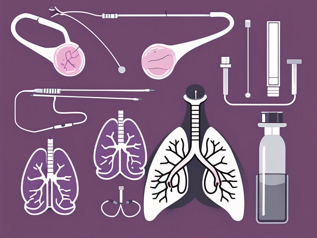
Bronchoscopy Biopsy
During a bronchoscopy, a thin, flexible tube (bronchoscope) is passed through your nose or mouth into your lungs. This allows the doctor to look directly inside the airways and collect small tissue samples from suspicious areas. This procedure is especially useful if the tumor is accessible through the airways.
Needle Biopsy
Also called a percutaneous biopsy, this method uses a thin needle inserted through the chest wall to reach lung tissue. Imaging tools like CT scans or ultrasound guide the needle to the exact spot. Needle biopsy is less invasive, performed on an outpatient basis, and commonly used if the lung abnormality is located closer to the chest surface.
Surgical Biopsy
When smaller biopsies don’t provide enough tissue or when the tumor is hard to reach, a surgical biopsy may be necessary. This involves making a small incision in the chest under general anesthesia to remove a piece of lung tissue. It’s performed in a hospital and allows a more thorough examination.
Your medical team will recommend the best option based on your tumor’s location, size, and your overall health. To learn more about biopsy techniques, Mayo Clinic offers an insightful overview on their lung biopsy page.
How Imaging Helps Guide Your Biopsy
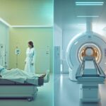
Imaging techniques like CT scans and ultrasound make biopsies safer and more precise. They provide real-time pictures, helping doctors navigate to suspicious areas and avoid critical structures in the lungs.
- CT-guided Needle Biopsy: If the tumor is deep or in a tricky area, CT imaging helps the doctor guide the needle accurately during the biopsy.
- Ultrasound Guidance: Uses sound waves to visualize the area and needle placement, commonly used for lesions near the chest surface.
Accurate targeting reduces risks and increases the chance of obtaining a representative tissue sample, which is crucial for an accurate diagnosis.
What to Expect During Your Lung Tissue Biopsy
Going through a biopsy might feel intimidating, but knowing the typical steps can ease your mind:
- Preparation: Your doctor will give you specific instructions, such as fasting or adjusting medications. Make sure to discuss any allergies or health conditions you have.
- Anesthesia: Depending on the biopsy type, you may receive local anesthesia to numb the area or general anesthesia to keep you asleep and comfortable.
- The Biopsy Procedure: Whether through bronchoscopy, needle, or surgery, the doctor will carefully remove a tissue sample from the area of concern.
- Recovery: Afterward, you’ll be monitored for any immediate side effects like bleeding or breathing difficulties.
- Sample Analysis: The tissue is examined by pathologists, which can take several days.
- Discussing Results and Next Steps: Your doctor will review the findings with you and discuss treatment options tailored to your diagnosis.
If you want a closer look at what a bronchoscopy entails, the Mayo Clinic’s patient education videos are a trusted resource.
Understanding Risks and Benefits
Though generally safe, biopsy procedures carry some risks, including:
- Minor bleeding or bruising
- Infection at the biopsy site
- Injury to surrounding lung tissue
Your care team will take every precaution to minimize these risks. Remember, getting an accurate diagnosis through biopsy gives you the best chance for timely, effective treatment and a better outcome.
Frequently Asked Questions
- How long does it take to get biopsy results?
- Biopsy results usually take a few days to one week, depending on the complexity of the analysis. Your doctor will keep you informed throughout the process.
- Will the biopsy hurt?
- Local anesthesia is used to numb the biopsy area, so you should feel little to no pain during the procedure. You may experience some discomfort or soreness afterward.
- Can a biopsy confirm the stage of lung cancer?
- Yes. By examining the tissue, pathologists can identify the cancer type and provide information about its stage, which guides treatment decisions.
- What happens after a lung cancer diagnosis via biopsy?
- Your doctor will discuss treatment options with you, which may include surgery, chemotherapy, radiation, targeted therapy, or a combination, depending on your diagnosis.
- Are there less invasive alternatives to biopsy?
- Imaging tests alone cannot confirm lung cancer, so biopsy remains the gold standard for diagnosis. Some new techniques, like liquid biopsies, are emerging but are not yet a complete replacement.